2. How Do Heart Look And Where It Is Present?
Everybody has a different-sized heart. Adult hearts are approximately the size of two clenched fists, but
children’s hearts are approximately the size of one clenched fist. Heart proportions are 12 cm long, 8.5
cm wide, and 6 cm thick. The average weight of a human heart is 230–280 g for women and 280–340 g for
men, though these numbers can vary widely depending on the individual.
The heart sits atop the diaphragm and is protected by the thoracic cavity located in the front of the
chest, behind the sternum and coastal cartilage. It is found between the two lungs of your body, which
occupy the left-angled lateral area known as pleural cavities. It is present to the left of the sternum.
3. Understanding About The Parts Of Heart…
Pericardium-
Your heart is surrounded by a double-layered tissue called pericardium. The pericardial cavity can be
thought of as the space between the two layers. The pericardial fluid contained within this space serves
to cushion the heart during contractions and prevent damage from occurring from movement or impact.
The pericardium controls heart movement. It lubricates, protects, and prevents acute volume overload in
the heart.
What are the layers of the heart?
The different layers of the heart protect your heart, and restricts the expansion of the heart when your
blood volume increases.
Three heart tissue layers are:
Epicardium
The outermost layer, known as the epicardium, is composed of a combination of mesothelial cells, adipose
tissue, and elastic connective tissue.
The outer layer of the pericardium covers the roots of your heart’s main blood veins and is attached by
ligaments to the spinal cord, diaphragm, and other body components.
Myocardium
Myocardium is the middle layer of the heart’s muscle, also known as cardiac muscle. This tissue within
the heart wall contracts.
It is composed of cardiac muscles in a spiral arrangement of myocardium that squeeze blood through the
heart and into the blood vessels in the correct orientations.
It is the thickest of the three layers and contains numerous mitochondria that provide energy to the
heart’s muscle cells.
Endocardium
The endocardium lines the innermost and deepest part of the heart. It’s a thin, smooth layer of tissue
that shields your heart’s chambers and valves from damage.
The inner layer of heart walls contains
the required blood vessels and functions as a barrier between the heart muscles and the blood
circulation.
The cardiac conduction system controls the electrical impulses sent between the heart’s
muscle cells.
4. About Chambers Of Heart-
Auricles- Atria are the two upper heart chambers. Thin-walled chambers receive blood from your veins. The
left atrium receives oxygen-rich blood from the lungs, whereas the right atrium receives deoxygenated
blood from the lower and upper bodies.
Right atrium is the heart’s upper chamber. The right atrium
receives deoxygenated blood from the body during a cardiac cycle. When both atria are full, they
contract and deoxygenated blood from the right atrium enters the right ventricle through the open
triangle valve
Left atrium is the upper left cardiac chamber. Pulmonary veins carry oxygenated blood to the left atrium
during a typical cardiac cycle. When full, both atria contract and oxygenated blood from the left atrium
flows through the open mitral valve into the left ventricle.
Ventricles- The ventricles are the two chambers at the bottom of the heart. The two ventricles are
chambers with thick walls that push blood out of the heart and into the lungs and other parts of your
body.
Right ventricle is lower right heart chamber. When the right atrium contracts, deoxygenated blood enters
the right ventricle. Right atrium valve closure fills right ventricle. Both ventricles constrict when
full. Lungs receive right ventricle blood.
Left ventricle is the lower left cardiac chamber. When the left ventricle contracts, the mitral valve
delivers oxygenated blood from the left atrium. The mitral valve closes and the aortic valve opens when
the left ventricle contracts. Closing the mitral valve keeps blood from returning to the left atrium,
while opening the aortic valve lets blood flow into the aorta and throughout the body.
5. What Is Heart Valves?
The heart valves serve as gateways at the chamber entrances, opening and shutting to allow blood flow.
Their fundamental responsibility is to ensure that blood flows in only one direction through the heart.
The valves are well designed to tolerate any physical impact, including high blood pressure and
velocity.The heart valves serve as gateways at the chamber entrances, opening and shutting to allow
blood flow. Their fundamental responsibility is to ensure that blood flows in only one direction through
the heart. The valves are well designed to tolerate any physical impact, including high blood pressure
and velocity.
Heart mainly has 4 major valves:
Tricuspid Valve
The opening of the tricuspid valve facilitates the flow of blood from the right atrium to the right
ventricles.
Mitral/Bicuspid Valve
Mitral valve opening allows blood to flow from the left atrium to the left ventricle. The mitral valve
allows oxygen-rich blood from the lungs to enter the left ventricle from the left atrium.
Aortic Valve
The aortic valve is the final valve through which oxygen-rich blood travels before leaving the heart and
circulating throughout the body. The valve inhibits the return of blood to the left ventricle.
Pulmonary Valve
The pulmonic or pulmonary valve is the valve through which deoxygenated blood flows. It restricts the
right ventricle and opens the pulmonary artery to allow blood flow to the lungs.
6. What Do You Mean By Heart Sound?
The valves of the heart open and close simultaneously to allow blood flow. The rapid closure of the
valves causes the surrounding fluid to vibrate and creates heart sound.
They are of two types:
Lub– The first cardiac sound, or S1, is generated by the closure of the atrioventricular valves
(mitral and tricuspid) at the start of ventricular contraction, or systole.
7. What Is The Significance Of Heart Sound?
It is valuable first line in patient’s evaluation. Abnormal heart sounds may be indicative of valve
related heart problems. Murmurs are the sound produced by the backflow of blood due to ineffective
valves.
8. Functioning And Working Of Heart?
The heart’s primary function is to maintain a steady blood flow throughout the body. Hence, the cells and
tissues are supplied with oxygen and nutrients.
Significant functions of heart:
It regulates the rhythm and rate of your heartbeat.
It regulates the rhythm and rate of your heartbeat.
The heart is also responsible for maintaining a healthy blood pressure level.
There are two types of circulation within the body: pulmonary circulation and systemic circulation heart
facilitates in both.
In cooperation with other body systems, your heart regulates your heart rate and other bodily functions.
9. Steps Involving Blood Flow-
The heart pumps oxygen- and nutrient-rich blood throughout your body with each beat. When blood returns
to the heart, it is sent to the lungs for oxygenation. The heart subsequently pumps blood to the rest of
the body, and the cycle repeats.
Blood enters the right atrium from the superior and inferior vena cava.
Blood from the right atrium flows through the right AV valve (tricuspid) into the right ventricle.
Contraction of the right ventricle forces the pulmonary semilunar valve to open.
Blood flows through the pulmonary valve into the pulmonary body.
Blood is distributed through the right and left pulmonary arteries to the lungs, where CO2 leaves and
loads oxygen.
Blood returns from the lungs to the left atrium via the pulmonary veins.
Blood from the left atrium flows through the left AV (mitral) valve into the left ventricle.
Contraction of the left ventricle forces the aortic semilunar valve to open.
Blood flows through the aortic valve into the ascending aorta.
Blood from the aorta is distributed to every organ of the body, where it expels O and charges CO2.
Blood returns to the heart from the vena cava.
10. Pacemakers-
Pacemakers are the heart tissues involved in pulse generation and heartbeat conduction.
Pacemaker tissue mainly consist of following basic parts:
Sino Atrial Node (SA Node)
Atrioventricular node (AV Node)
Atrioventricular Bundle/ Bundle of His (AV Bundle)
Purkinje Fibres
11. SA Node (Sino Atrial Node)
The SA node is responsible for maintaining a proper heart rate and rhythm by continuously producing
electrical impulses. Thus, the SA node is known as the heart’s natural pacemaker. SA node is a thin,
elongated, glycogen- and mitochondria-rich muscle fiber. It is situated near the confluence of the
superior vena cava and right atrium. Generating electrical impulses at regular intervals to establish
and sustain a steady heartbeat. SA node is also involved in heart rate regulation.
12. AV Node (Atrioventricular Node)
It is regarded to be the heart’s secondary pacemaker. The AV node is a mass of specialized cells capable
of self-excitation. Its principal role is impulse transmission to the ventricles. It can generate
electrical impulses and conducts them from the heart’s atria to ventricles.
13. AV Bundle (Atrioventricular Bundle/Bundle Of His)
Bundle of his commences at the AV node, which is a group of fibers that send electrical impulses through
the center of the heart. The bundle of His is an extended segment connecting the AV node with the left
and right bundle branches of the septal crest. The bundle of His is composed of a right and left branch.
In the ventricles, both branches divide frequently to form a network of fibers.
14. Purkinje Fibers
Purkinje fibers come from the terminal divisions of the right and left His bundle branches to pierce the
ventricular wall. They are greater in size and density than the SA node. Purkinje fibers have a big
diameter and send impulses at a rapid rate.
15. How To Keep Your Heart Healthy?
Maintaining a healthy heart is crucial for excellent overall health and minimizing the risk of heart
disease and other cardiovascular issues. Here are some heart-healthy practices:
Healthy Diet: Consume a diet rich in fruits, vegetables, whole grains, lean proteins, and healthy fats.
Limit your consumption of saturated and trans fats, sugars added to foods, and salt.
Exercise frequently: Strive for at least 30 minutes of moderate-intensity activity on most days of the
week. Examples include brisk walking, cycling, swimming, and dancing.
Keep a healthy weight: Being overweight or obese increases the risk of heart disease. Aim to reduce
weight through a combination of good nutrition and activity if you are overweight.
Don’t smoke: Smoking is a big risk factor for cardiovascular disease and can cause damage to your blood
vessels and heart. If you smoke, seek help to quit.
Stress management is important since chronic stress can lead to high blood pressure and other heart
issues. Discover methods of stress management, such as meditation, yoga, and deep breathing.
Obtain sufficient rest: Strive for 7-9 hours of sleep per night. Sleep deprivation can increase the risk
of hypertension, obesity, and other cardiac diseases.
Keep an eye on your blood pressure and cholesterol levels. Excessive blood pressure and cholesterol
might raise the risk of heart disease. Periodically check these levels and, if required, consult with
your physician to manage them.
By adhering to these steps, you can maintain a healthy heart and lower your risk of heart disease and
other cardiovascular complications.
Other Heart blogs
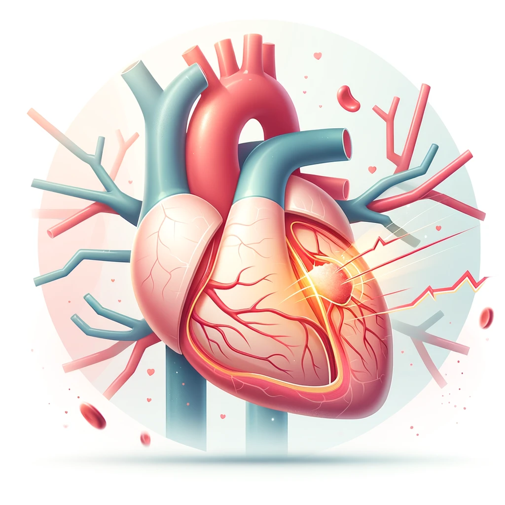
Heart Attack
Myocardial infarction (=heart attack) is an exceedingly severe disorder that occurs when the
heart muscle is deprived of blood supply. Without blood flow, the afflicted heart muscle will
begin to die. A heart attack can result in irreversible damage to the heart and...
learn more
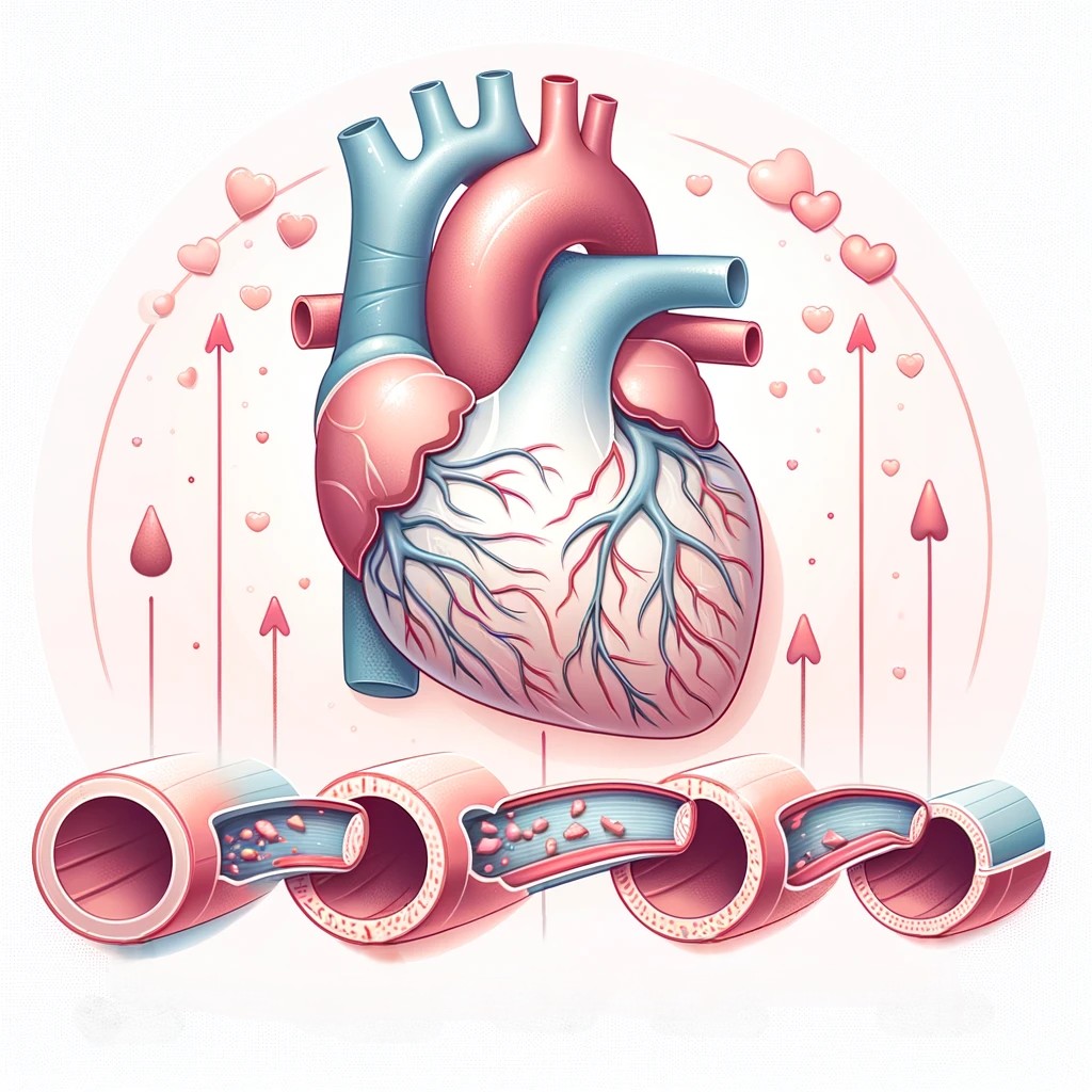
Coronary Artery Disease
Coronary artery disease is a prevalent heart disorder that affects the primary blood arteries
that feed
the heart muscle with blood. CAD can result in a heart attack or other consequences, including
arrhythmia and heart failure. With coronary artery disease...
learn more
Heart Transplant
A heart transplant is a surgery in which your heart is replaced with a donated heart from another
person. Heart transplantation is typically reserved for patients whose condition has not
improved sufficiently with medications or other surgical procedures...
learn more
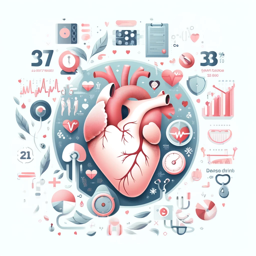
Heart Disease Facts
In India, heart disease is a major public health concern and the leading cause of death. Here are
some heart disease statistics from India:
Cardiovascular diseases (CVDs) are the leading cause of death in
India, accounting for 28% of all deaths, according to the World Health Organization.
In India, the age-standardized death rate from CVDs is 272 per
100,000 people.
...
learn more